
Excellent work, your patient tolerated the kidney biopsy well. Let’s check out the histology below. Click here for some quick pathology tips.
Images courtesy of Dr. Tiffany Caza, Arkana Laboratories

First things first – what’s that stain?
Methenamine Silver
Nice! Notice that the tubules and capillary loops appear black, as the JMS stains collagen black.
Periodic-Acid Schiff (PAS)
Try again!
Hematoxylin & eosin (H&E)
Pick again!
Masson’s Trichrome
Try again!
Immunohistochemistry (IHC)
Give it another shot!
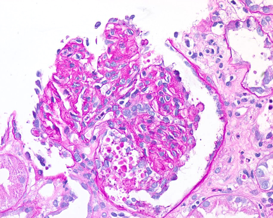
How would you describe the glomerular injury pattern seen above?
Collapsing focal segmental glomerulosclerosis (FSGS)
Strong work! We see collapse of the capillary loops as well as hypertrophy and hyperplasia of overlying podocytes outside the capillary loops. Below is a periodic acid-Schiff (PAS) slide from this patient showing the same pattern, as well as a normal glomerulus (methenamine silver stain)
 Normal glomerulus (Jones silver)

 Normal glomerulus (Jones silver)
Focal segmental glomerulosclerosis, tip lesion variant
Close – but we don’t see a lesion here involving the tip domain (the outer part of the tuft next to the origin of the proximal tubule). Try again!
Minimal change disease (MCD)
Remember that MCD is named because of minimal changes on light microscopy. This glomerulus looks quite abnormal! Choose again!
Membranoproliferative glomerulonephritis (MPGN)
We’re not seeing the lobular appearance of the glomerulus here. On a JMS stain, we may also be able to visualize duplication of the basement membrane (“tram-tracks”) – which are not visible here. Give it another shot!
Subepithelial deposits
Though deposits are better visualized under electron microscopy, a “spike and hole” pattern may be seen on the methenamine silver stain in individuals with membranous nephropathy, as the stain will leave deposits unstained (hole) between “spikes” of black-stained basement membrane (see below). We don’t see that here…try again! 

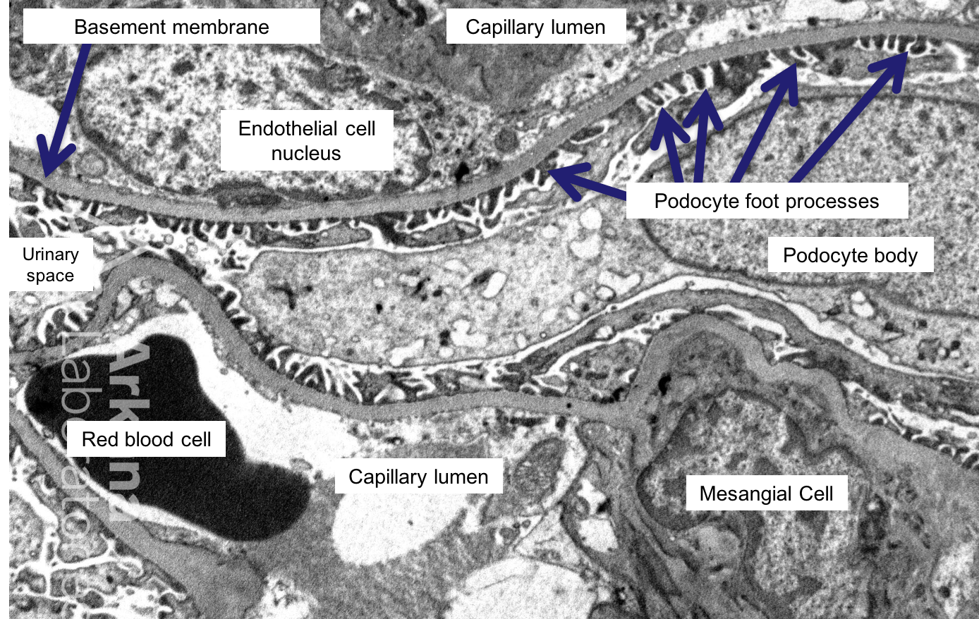
Here’s an electron micrograph (EM) from this patient…what do you see?


Podocyte foot process effacement
Nice! Yes, the podocytes are flattened/effaced here. Note the difference above, when compared to the normal EM below:

Sub-epithelial deposits
We don’t see electron dense (i.e. dark) deposits here. Try again!
Sub-endothelial deposits
We don’t see electron dense (i.e. dark) deposits here. Try again!
Mesangial deposits
We don’t see electron dense (i.e. dark) deposits here. Try again!
Basement membrane thickening
The glomerular basement membrane appears to be a normal thickness here. Pick again!
Polymorphisms or mutations in which of the following genes is most likely to be associated with the diagnosis here?
Apolipoprotein L1 (APOL1)
Nailed it!
Podocin (NPHS2)
Individuals with a mutation in this gene are more likely to present at a younger age. Choose again!
Nephrin (NPHS1)
Individuals with a mutation in this gene are more likely to present at a younger age. Try again!
Polycystin-1 (PKD1)
Mutations in this gene are associated with polycystic kidney disease. Pick again!
Nephrocystin 1 (NPHP1)
Mutations in this gene are associated with nephronophthisis. Pick again!
Click here to wrap up this case!
Case 50 Index
Case 50 Introduction
Case 50 Physical Exam
Case 50 Diagnostic Testing
Case 50 Additional Diagnostic Testing