
Welcome to the Urine Gallery!
Click on the images below to enlarge them.
Thank you to Dr. Jay Seltzer and Jose Poloni for sharing these images.
Ready to spin some urine? Check out our “How to Spin Urine” guide here. To learn more about different urine microscopy elements, visit Renal Fellow Network’s “Urine Sediment of the Month” series. #SpinUrine #UrineMicroscopy Abbreviations: Epi = epithelial WBC = white blood cells RBC = red blood cells.
Here’s a NephSIM Urine Microscopy JEOPARDY! game board.
Check out Dr. Jay Seltzer’s KIDNEYcon 2022 Urine Microscopy video here with dazzling images!
Images are organized in the sections below:
- Unstained Casts & Acanthocytes
- Stained Casts & Acanthocytes
- Phase Contrast Casts & Acanthocytes
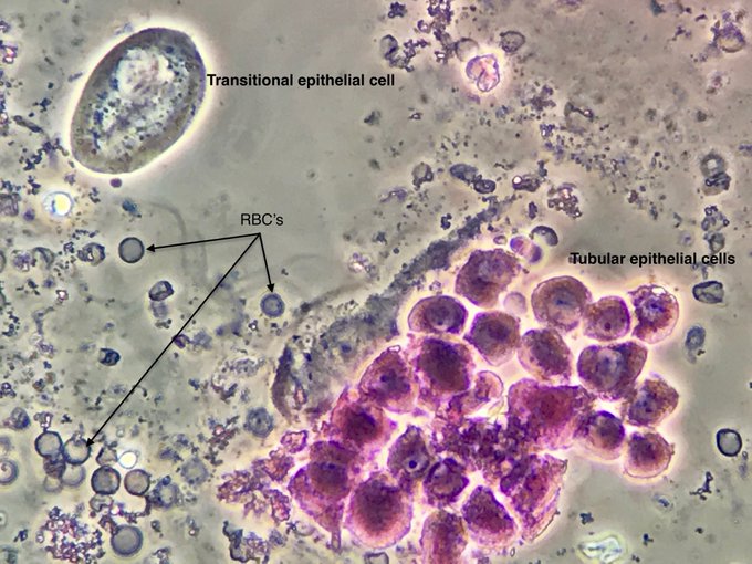
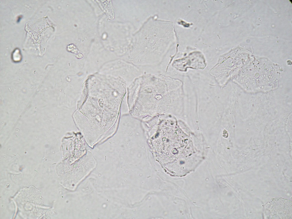
- Squamous, Transitional, and Renal Tubular Epithelial Cells
- Lipids
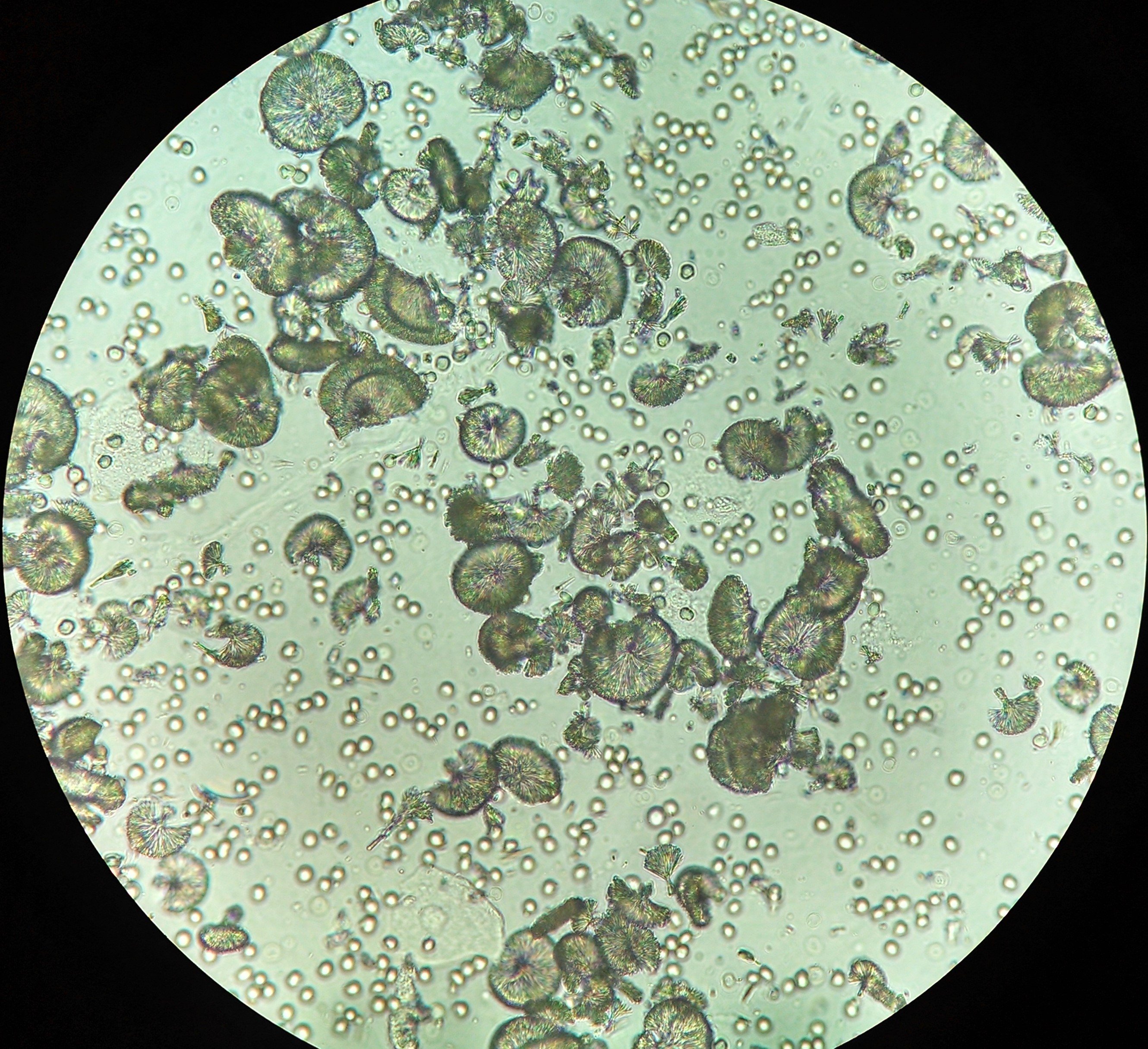
- Crystals
- Other
Unstained Casts & Acanthocytes (Brightfield Illumination; Images Courtesy of: Dr. Jay Seltzer and Jose Poloni )


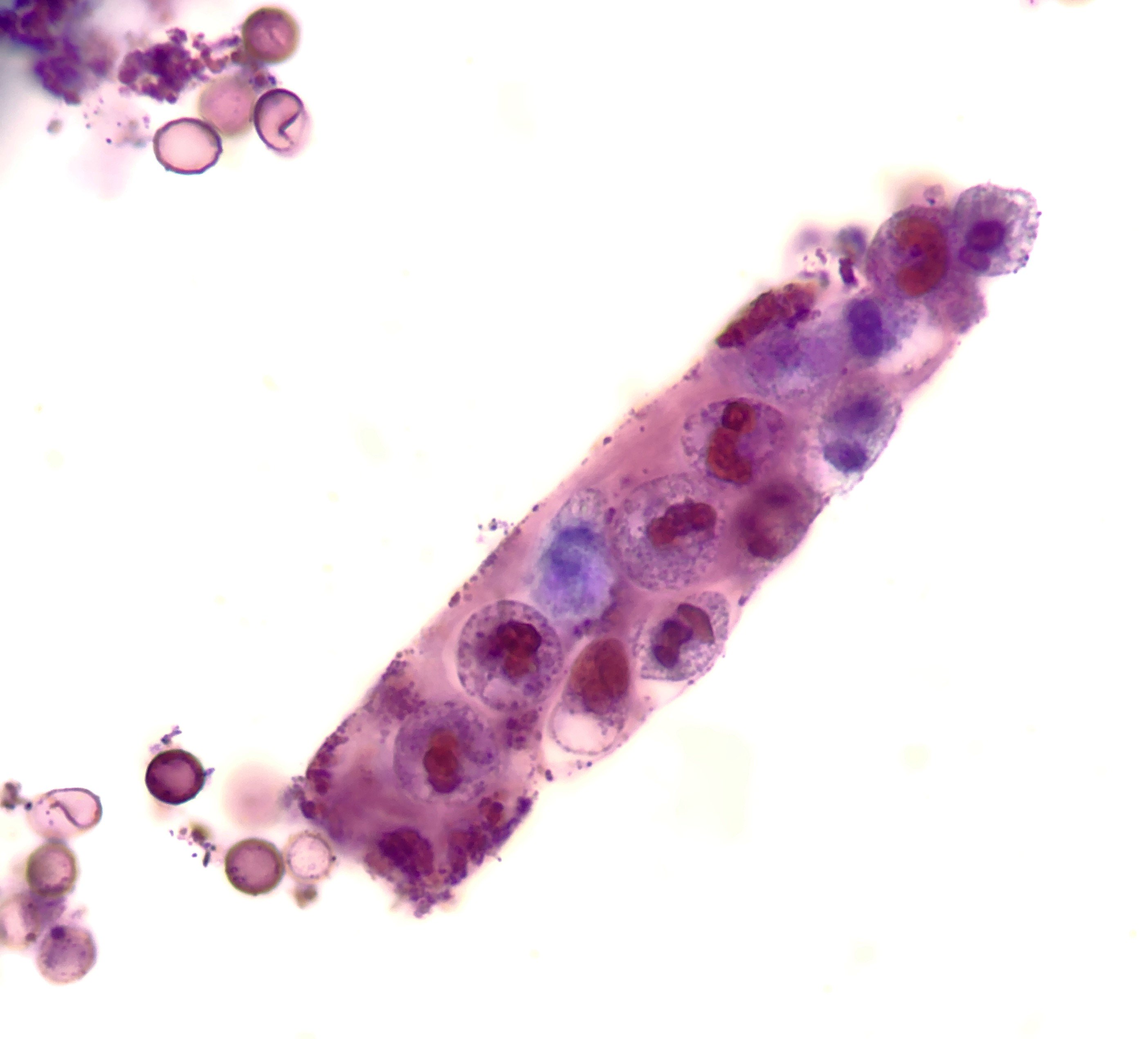
Stained Casts & Acanthocytes (Sternheimer-Malbin Stain, Brightfield Illumination; Images Courtesy of Dr. Jay Seltzer)


Phase Contrast Casts & Acanthocytes (Images Courtesy of Dr. Jay Seltzer; SM = Sternheimer Malbin stain)


Squamous, Transitional, and Renal Tubular Epithelial Cells (Images Courtesy of Dr. Jay Seltzer; RTEC = Renal Tubular Epithelial Cells; SM = Sternheimer Malbin stain)


Lipids (Images Courtesy of Dr. Jay Seltzer; SM: Sternheimer Malbin Stain)


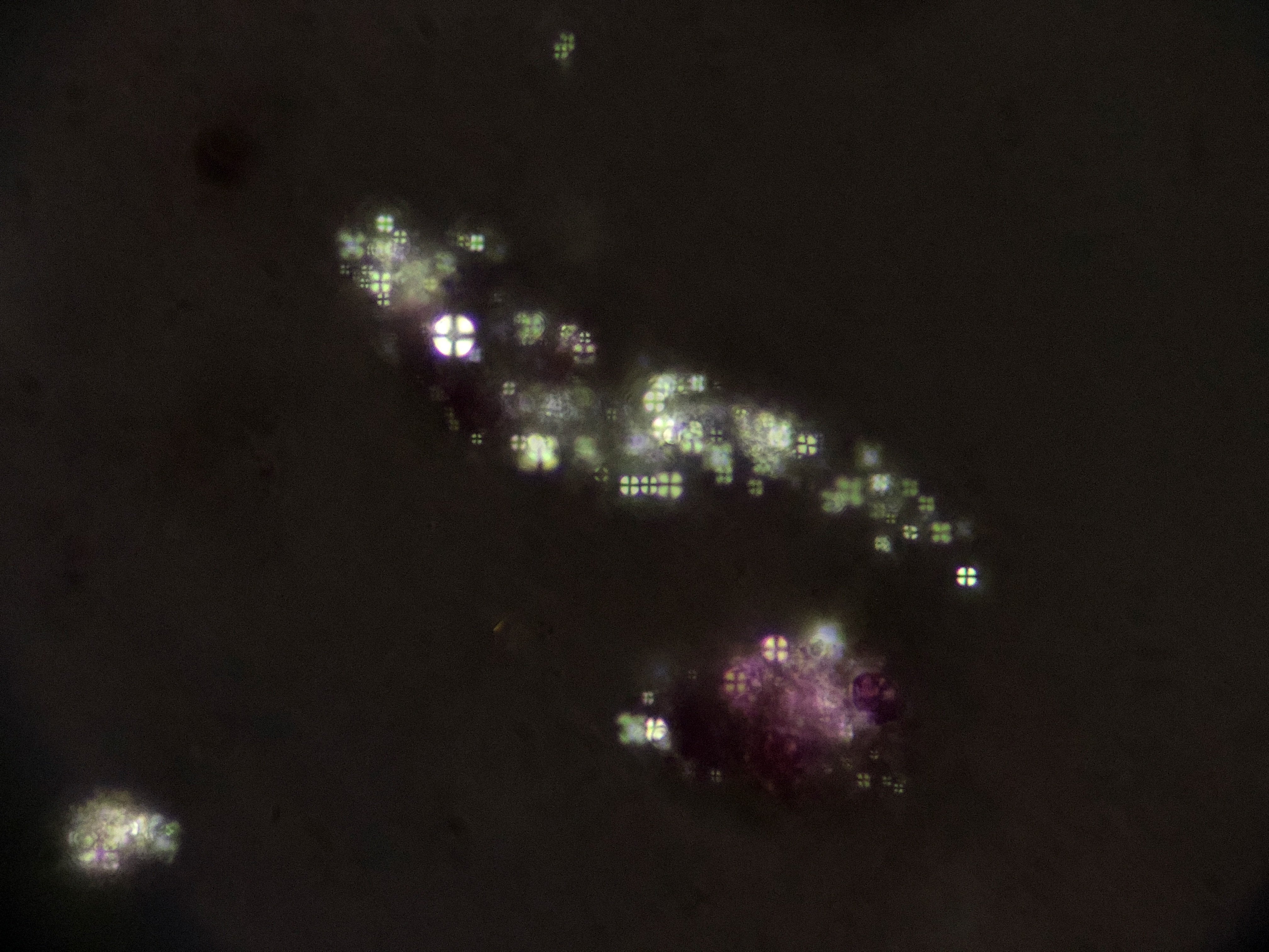


Crystals (Images Courtesy of Jose Tesser Poloni & Dr. Jay Seltzer)


Other (Images Courtesy of Dr. Jay Seltzer; SM: Sternheimer Malbin Stain)


Urine Microscopy Cells & Casts

Stones & Crystals

Urine Microscopy Cartoons (Courtesy of Dr. Krystahl Andújar, @Krystahllopathy)


