
Your pathologist informs you the additional slide preparations are ready! Take a look below…
EM (Electron Microscopy)

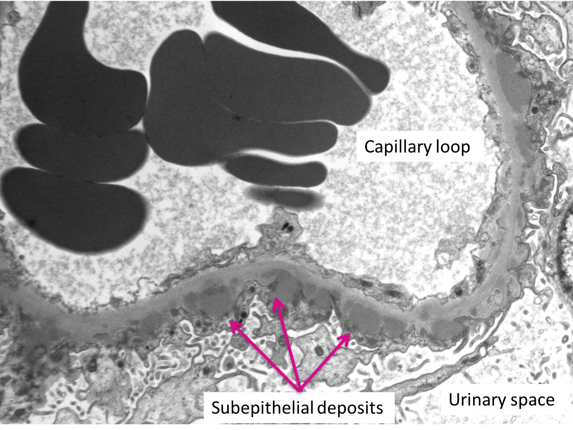
Where do you see the majority of deposits in the electron microscopy above?
There are no deposits.
Try again! We see dark gray, dense material (deposits) in the electron micrograph above.
Mesangial deposits
We don’t actually see the mesangial space here and thus cannot identify mesangial deposits. Pick again!
Subendothelial deposits
The majority of these deposits are not in the subendothelial space, take another look!
Subepithelial deposits
Correct!

Here’s another stain:

What stain do you see above?
Jones Methamine Silver Stain
Correct! Collagen appears black with this stain. In this disease process, we see the “spike and hole appearance.” The “spikes” of basement membrane are seen as black, while the deposits (“holes”) do not pick up the stain. These “holes” may give the basement membrane a “bubbly” appearance.
Periodic Acid-Schiff (PAS) Stain
In a PAS stain, collagen appears magenta. Pick again!
Immunohistochemistry (IHC) stain for PLA2R
This is not an IHC stain. When an IHC stain is positive, the antigen of interest should appear brown. Try again!
Masson’s Trichrome Stain
This stain is used to visualize fibrosis. The colors in a Masson’s Trichrome stained slide should appear tones of red and blue. Pick again!
We’re almost there! Take a look at the immunofluorescence stains below (click!)
IgG 

IgA 
PLA2R

C3
C1q
Click here to confirm your diagnosis!
Case 35 Index
Case 35 Introduction
Case 35 Physical Exam
Case 35 Diagnostic Testing
Case 35 Pathology