
Great job, your patient tolerated the kidney biopsy well. The next day, your pathologist shows you the H&E stain below. Click here for some quick pathology tips.
What do you see below?
Podocyte effacement
With an H&E stain, it can be difficult to visualize the podocytes. We would need electron microscopy to assess the podocytes. Choose again!
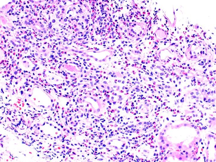
Interstitial infiltration with inflammatory cells
Correct! Click below to move on!
Subendothelial deposits
We cannot visualize deposits in the basement membrane with an H&E stain. Electron microscopy is typically needed to look for deposits. Take another shot!
Endocapillary cell proliferation with obliteration of capillary loops and mesangial matrix expansion
Both capillary loops and mesangial cells are found within the glomerulus, and your pathologist hasn’t shown you a glomerulus below. Try again!


Your pathologist tells you other stains will take 1-2 more days.
Click here when you’re ready to look at more stains that might help you confirm your diagnosis!
Case 2 Index
Case 2 Introduction
Case 2 Physical Exam
Case 2 Diagnostic Testing